



























What Is a Chest Tube? A Comprehensive Guide to Its Applications, Types, and Role in Respiratory and Surgical Care
Introduction
The respiratory system is one of the most vital systems of the human body, and the proper functioning of the lungs is essential for delivering oxygen to body tissues and sustaining life. For normal respiration, the lungs require adequate space within the thoracic cavity, known as the pleural space. Any abnormal accumulation of air, blood, or fluid within this space can impair normal lung function and, in certain cases, create a life-threatening medical emergency.
In many clinical conditions—including pneumothorax, hemothorax, pleural effusion, pleural infections, and following various cardiac and thoracic surgical procedures—the removal of abnormal contents from the pleural cavity becomes necessary. One of the most important medical devices used for this purpose is the chest tube, also known as a thoracic drainage tube.
A chest tube creates a controlled pathway for the evacuation of excess air and fluids, helping restore the normal negative pressure within the pleural cavity, allowing the lung to re-expand, and improving the patient’s respiratory function. The tube is typically connected to a chest drainage system, which regulates drainage, prevents the backflow of air or fluid, and ensures the safety and effectiveness of the treatment process.
Given the critical role of chest drainage systems in emergency medicine and postoperative care, selecting high-quality equipment, well-designed drainage units, and reliable collection systems is essential for improving patient safety and minimizing complications.
This article provides a comprehensive overview of chest tubes, including their indications, types, mechanisms of action, components of chest drainage systems, characteristics of standard drainage equipment, and the importance of using safe and reliable systems in healthcare facilities.
What Is a Chest Tube?
A chest tube is a sterile, flexible medical tube inserted through the chest wall into the pleural space to remove abnormal accumulations of air, blood, fluids, or other pathological secretions.
The procedure, commonly known as tube thoracostomy or chest drain insertion, is one of the most frequently performed interventions in thoracic medicine and is carried out by trained healthcare professionals under various clinical circumstances.
Chest tubes are typically manufactured from flexible, biocompatible medical-grade polymers that provide sufficient flexibility while maintaining resistance against kinking, obstruction, and mechanical damage.
A complete chest drainage system generally consists of the following components:
- Chest Tube: The primary conduit for draining air and fluids from the pleural cavity.
- Drainage Catheter: Connects the chest tube to the drainage unit and transports the collected contents.
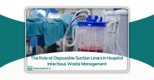
- Chest Drainage Unit (CDU): The collection device that safely stores drained fluids.
- Water Seal Chamber: Prevents air from flowing back into the pleural cavity while allowing air to escape.
- Suction Control Chamber: Regulates negative pressure in systems that utilize suction.
Together, these components create a closed, controlled drainage system that enables the safe evacuation of pleural contents while minimizing the risk of contamination and complications.
Understanding the Pleural Space and Its Importance
To better understand the role of a chest tube, it is essential to first understand the anatomy and function of the pleural space.
The lungs are enclosed by two thin membranes collectively known as the pleura:
- Visceral Pleura: The inner membrane that directly covers the surface of the lungs.
- Parietal Pleura: The outer membrane lining the inner surface of the chest wall.
Between these two layers lies a narrow space called the pleural space (or pleural cavity). Under normal physiological conditions, this space contains a small amount of lubricating pleural fluid, which reduces friction as the lungs expand and contract during breathing.
More importantly, the pleural space maintains a negative intrapleural pressure, which is essential for keeping the lungs fully expanded.
However, this delicate balance can be disrupted by the accumulation of air or fluid.
For example:
- Air entering the pleural cavity results in pneumothorax.
- Blood accumulation leads to hemothorax.
- Excess fluid causes pleural effusion.
These conditions increase pressure within the pleural cavity, compress the lung, impair gas exchange, and reduce respiratory efficiency. A chest tube removes these abnormal contents, allowing the lung to re-expand and restoring normal respiratory mechanics.
Why Is a Chest Tube Necessary?
The primary purpose of chest tube insertion is to establish a safe pathway for removing abnormal air or fluid from the pleural cavity. Without adequate drainage, the accumulation of these substances may lead to severe respiratory compromise and, in some cases, life-threatening complications.
The main objectives of chest tube therapy include:
1. Restoring Normal Pleural Pressure
Excess air or fluid disrupts the negative pressure required for normal lung expansion. By evacuating these substances, the chest tube helps restore physiological pleural pressure and allows the lung to re-expand.
2. Improving Respiratory Function
When the lung is compressed, its ability to exchange oxygen and carbon dioxide is significantly reduced. Effective pleural drainage improves ventilation, enhances oxygenation, and relieves respiratory distress.
3. Preventing Reaccumulation of Air or Fluid
In many patients, air leakage or fluid production continues after the initial diagnosis or surgery. Chest drainage systems provide continuous and controlled evacuation, preventing recurrent accumulation.
4. Postoperative Management
Following cardiac, pulmonary, and thoracic surgical procedures, blood, air, and serous fluids may accumulate within the chest cavity. Chest tubes play a crucial role in postoperative care by preventing these collections and reducing the risk of complications.
Primary Clinical Applications of Chest Tubes
Chest tubes are among the most commonly used devices in emergency medicine, thoracic surgery, intensive care, and postoperative patient management. Their primary function is to evacuate air, blood, and other abnormal fluids from the pleural cavity, thereby restoring normal pulmonary function.
Depending on the patient’s condition, chest tubes may be indicated in several clinical situations.
1. Pneumothorax
Pneumothorax occurs when air enters the pleural cavity, disrupting the normal negative intrapleural pressure and causing partial or complete lung collapse.
Common causes include:
- Penetrating or blunt chest trauma
- Rib fractures
- Spontaneous rupture of pulmonary blebs
- Chronic lung diseases such as Chronic Obstructive Pulmonary Disease (COPD)
- Complications of certain medical procedures
In these cases, the chest tube removes the trapped air, allowing the lung to re-expand and resume normal function.
2. Hemothorax
Hemothorax refers to the accumulation of blood within the pleural cavity.
It commonly results from:
- Major trauma
- Chest injuries
- Cardiac or thoracic surgery
- Vascular injury
Blood accumulation not only compresses the lung but may also contribute to significant blood loss and hemodynamic instability. Chest tube drainage enables blood evacuation, continuous monitoring of bleeding volume, and assists clinicians in making appropriate treatment decisions.
3. Pleural Effusion
Several medical conditions can cause excessive fluid accumulation within the pleural space.
Common causes include:
- Congestive heart failure
- Lung and pleural malignancies
- Liver disease
- Kidney disease
- Inflammatory disorders
Large pleural effusions may result in shortness of breath and reduced lung capacity. In these cases, chest tubes can provide continuous drainage to relieve symptoms and improve respiratory function.
4. Chylothorax
Chylothorax is characterized by the accumulation of lymphatic fluid (chyle) within the pleural cavity.
Potential causes include:
- Injury to the thoracic duct
- Thoracic surgical procedures
- Certain malignancies
Chest tube drainage is commonly used to remove chyle and facilitate patient recovery.
5. Postoperative Cardiac and Thoracic Surgery
Nearly all patients undergoing major thoracic procedures require chest tubes after surgery.
Common examples include:
- Open-heart surgery
- Coronary artery bypass grafting (CABG)
- Heart valve replacement
- Lobectomy
- Pneumonectomy
- Video-Assisted Thoracoscopic Surgery (VATS)
In these patients, chest tubes continuously remove blood, air, and postoperative secretions, preventing pressure buildup around the lungs and heart while promoting safe recovery.
Types of Chest Tubes
Chest tubes are available in various sizes, designs, and configurations to accommodate different clinical indications. The selection of an appropriate chest tube depends on the patient’s condition, the nature of the pleural pathology, and the physician’s clinical judgment.
Classification by Size (French Scale)
Chest tube diameter is typically expressed using the French (Fr) scale.
In general:
- 10–14 Fr: Commonly used for pneumothorax and small-volume pleural effusions.
- 16–24 Fr: Suitable for most routine pleural drainage procedures.
- 28–36 Fr: Recommended for hemothorax and significant blood drainage.
- 36–40 Fr: Primarily used after major cardiac and thoracic surgeries.
Selecting the correct tube size is essential to ensure effective drainage while minimizing patient discomfort and tissue trauma.
Classification by Design
Straight Chest Tube
The straight chest tube is the most widely used design in clinical practice.
It is suitable for evacuating both air and pleural fluid and is commonly employed in emergency medicine, thoracic surgery, and intensive care settings.
Right-Angle Chest Tube
A right-angle chest tube is primarily designed for postoperative cardiac surgery.
Its angled configuration allows optimal placement within the mediastinum, making it particularly effective for draining blood and fluid around the heart following surgical procedures.
Pigtail Catheter
The pigtail catheter is a small-bore catheter with a curled distal tip designed to minimize displacement after insertion.
Over recent years, pigtail catheters have become increasingly popular as an alternative to large-bore chest tubes in selected patients with uncomplicated pneumothorax or small pleural effusions.
Their advantages include:
- Reduced procedural pain
- Smaller incision size
- Improved patient comfort
- Less invasive insertion technique
How Does a Chest Drainage System Work?
A chest tube alone cannot safely evacuate air or fluids from the pleural cavity. To function effectively, it must be connected to a Chest Drainage System (CDS).
This drainage system regulates fluid collection, prevents air from re-entering the pleural cavity, and, when necessary, provides controlled suction to facilitate lung re-expansion.
A standard chest drainage system generally consists of three major components.
1. Collection Chamber
The collection chamber is designed to collect and safely store:
- Blood
- Pleural fluid
- Surgical drainage
- Other pathological secretions
Modern collection chambers include clearly graduated volume markings, allowing healthcare professionals to continuously monitor drainage output.
Accurate measurement of drainage volume is essential for:
- Detecting ongoing bleeding
- Monitoring postoperative recovery
- Evaluating treatment effectiveness
- Determining when chest tube removal is appropriate
2. Water Seal Chamber
The water seal chamber is considered the most critical component of the chest drainage system.
It functions as a one-way valve, allowing air to escape from the pleural cavity while preventing air from flowing back into the chest.
This mechanism is essential for maintaining negative intrapleural pressure and promoting lung re-expansion.
Observation of bubbling within the water seal chamber may indicate that air is escaping from the pleural cavity. In certain situations, persistent bubbling may suggest the presence of an air leak, which requires clinical evaluation.
3. Suction Control Chamber
In many patients, passive drainage alone is insufficient to achieve complete lung re-expansion.
In such cases, controlled negative pressure is applied through the suction control chamber.
The addition of suction can:
- Accelerate the removal of air and fluids
- Promote faster lung expansion
- Improve drainage efficiency
- Reduce postoperative complications
While traditional systems regulate suction using a water column, many modern devices employ dry suction technology, which offers greater precision, reduced noise, and easier operation.
Single-, Two-, and Three-Chamber Chest Drainage Systems
Chest drainage systems are commonly available in three basic configurations.
Single-Chamber System
The single-chamber system is the simplest design.
In this configuration, drainage collection and water seal functions are combined within a single chamber.
Although historically important, these systems are now rarely used because of their limitations in monitoring and safety.
Two-Chamber System
The two-chamber system consists of:
- A collection chamber
- A water seal chamber
This design remains widely used in many healthcare facilities and provides improved drainage monitoring compared with single-chamber systems.
Three-Chamber System
The three-chamber system represents the current standard of care in most modern hospitals.
It includes:
- Collection Chamber
- Water Seal Chamber
- Suction Control Chamber
This design provides superior drainage control, improved monitoring capabilities, and greater patient safety.
Today, the majority of tertiary hospitals and specialized thoracic surgery centers utilize three-chamber chest drainage systems.
The Role of the Chest Drainage Unit in Patient Safety and Treatment Success
Although the chest tube provides the pathway for evacuating air and fluids from the pleural cavity, the Chest Drainage Unit (CDU) is responsible for managing these materials safely and effectively.
The drainage unit is far more than a simple collection container. It is an integral component of patient care that performs several critical functions, including:
- Maintaining appropriate intrapleural pressure
- Preventing air reflux into the pleural cavity
- Measuring drainage volume
- Facilitating continuous monitoring of the patient’s clinical status
If the drainage system is poorly designed or manufactured with inadequate quality standards, complications such as air reflux, fluid leakage, impaired drainage, and an increased risk of infection may occur.
For this reason, selecting a high-quality, clinically validated chest drainage system is considered a fundamental requirement for patient safety in modern healthcare facilities.
Characteristics of a Standard Chest Drainage System
Advancements in medical technology have significantly improved modern chest drainage systems in terms of safety, accuracy, and ease of use. Compared with older models, today’s systems incorporate features that enhance clinical performance while reducing the risk of complications.
A high-quality chest drainage system should provide the following characteristics.
1. Completely Closed Chest Drainage System
One of the defining features of modern drainage systems is their closed-system design.
In a closed chest drainage system, air and fluids remain within a sealed pathway from the moment they leave the pleural cavity until final disposal.
The advantages of this design include:
- Reduced risk of environmental contamination
- Minimized exposure of healthcare workers to blood and body fluids
- Lower risk of pathogen transmission
- Improved safety for both patients and healthcare personnel
Closed drainage systems are now widely recommended in infection prevention and hospital safety guidelines.
2. Accurate Measurement of Drainage Volume
One of the most important clinical parameters following thoracic or cardiac surgery is the amount of fluid drained from the pleural cavity.
Therefore, the collection chamber should include clear and accurate graduated markings that allow healthcare providers to monitor drainage volume continuously.
Accurate drainage measurements are essential for:
- Assessing ongoing bleeding
- Detecting postoperative complications
- Determining the appropriate timing for chest tube removal
- Evaluating the patient’s recovery
3. Effective Anti-Reflux Mechanism
Backflow of air into the pleural cavity can interfere with lung re-expansion and delay recovery.
Standard chest drainage systems incorporate anti-reflux mechanisms that prevent reverse airflow while maintaining continuous drainage.
This feature ensures consistent system performance, even during patient movement or transportation.
4. Leak-Resistant Construction
Because chest drainage systems are continuously exposed to blood, pleural fluid, and other potentially infectious body fluids, the structural integrity of the collection unit is critically important.
High-quality medical-grade polymers provide excellent resistance to:
- Cracking
- Fracture
- Puncture
- Fluid leakage
A leak-resistant design protects both patients and healthcare personnel while maintaining the integrity of the drainage system.
5. Secure Connectors
All tubing connections should be designed to minimize the risk of accidental disconnection.
An unexpected interruption in the drainage pathway may lead to:
- Air entering the pleural cavity
- Reduced drainage efficiency
- Patient complications
- Delayed recovery
For this reason, connector quality is considered one of the key indicators of a reliable chest drainage system.
6. Easy-to-Read Chambers
In operating rooms, intensive care units, and emergency departments, rapid clinical assessment is essential.
An effective chest drainage unit should allow healthcare professionals to easily observe:
- Drainage volume
- Water seal fluctuations
- Presence of air leaks
- Suction performance
Clear visualization supports faster clinical decision-making and improves patient monitoring.
The Role of Chest Drainage Systems in Infection Prevention
Infection prevention is one of the primary objectives of modern chest drainage systems.
Earlier-generation systems were associated with a higher risk of blood or fluid leakage, increasing environmental contamination and occupational exposure among healthcare workers.
Contemporary closed drainage systems substantially reduce these risks through improved engineering and secure connections.
From an infection control perspective, high-quality chest drainage systems offer several important advantages:
- Reduced direct contact with blood and pleural secretions
- Lower risk of hospital environmental contamination
- Decreased transmission of infectious microorganisms
- Improved safety during fluid collection
- More efficient management of medical liquid waste
These benefits are particularly valuable in high-risk hospital departments such as:
- Intensive Care Units (ICUs)
- Coronary Care Units (CCUs)
- Operating Rooms
- Emergency Departments
Modern Technologies in Chest Drainage Systems
Continuous innovation has led manufacturers to develop chest drainage systems that are safer, more accurate, and easier to operate.
Several advanced technologies have become increasingly common.
Dry Suction Systems
Traditional chest drainage systems regulate suction pressure using a water column.
Modern Dry Suction Systems replace the water column with mechanical or electronic pressure regulators.
Their advantages include:
- Greater accuracy
- Reduced operating noise
- Simplified maintenance
- Lower risk of user error
- More consistent suction performance
As a result, dry suction technology has become the preferred option in many modern hospitals.
Digital Chest Drainage Systems
Some advanced drainage units incorporate digital monitoring technology capable of displaying:
- Air leak measurements
- Intrapleural pressure
- Drainage volume
- Treatment trends over time
These systems provide objective clinical data that support evidence-based decision-making and may contribute to earlier chest tube removal and shorter hospital stays.
Antibacterial Filters
Certain chest drainage systems include antibacterial filters designed to reduce contamination within the drainage pathway.
These filters help protect both the patient and the drainage equipment while minimizing the risk of microbial transmission.
Ergonomic Design
Modern chest drainage units are increasingly designed with usability in mind.
Common ergonomic improvements include:
- Easier transportation
- Smaller footprint
- Greater stability to reduce tipping
- Simplified setup and replacement
- Improved handling by healthcare personnel
These design enhancements improve workflow efficiency and reduce the likelihood of user-related errors.
Common Mistakes in Chest Tube Management
Even the most advanced chest drainage system cannot compensate for improper clinical practice.
Several common errors may compromise drainage efficiency and patient safety.
These include:
- Kinking or twisting of the chest tube, resulting in impaired drainage.
- Obstruction by blood clots, preventing effective evacuation.
- Positioning the drainage unit above the patient’s chest level, increasing the risk of fluid reflux.
- Accidental disconnection of tubing connections.
- Manipulating or emptying the system without maintaining sterile technique.
- Delayed assessment of drainage output.
- Failure to accurately document drainage volume and clinical observations.
Strict adherence to standardized clinical protocols, combined with ongoing staff education, significantly reduces the likelihood of these complications and improves patient outcomes.
How to Choose a Standard Chest Drainage System
Selecting an appropriate chest drainage system involves much more than choosing a collection container or a drainage tube. As an integral part of patient care, the quality and performance of the system can directly influence patient safety, clinical outcomes, and the efficiency of healthcare professionals.
When selecting a chest drainage system, healthcare facilities should evaluate several technical, safety, and quality criteria.
Medical-Grade Materials
The system should be manufactured from high-quality, biocompatible medical-grade polymers that provide excellent mechanical strength while maintaining compatibility with blood and other body fluids.
Premium materials reduce the likelihood of:
- Cracking
- Fluid leakage
- Structural deformation
- Mechanical failure
Completely Closed System
Closed drainage systems minimize healthcare workers’ exposure to blood and body fluids while reducing environmental contamination.
This feature is particularly important in:
- Operating rooms
- Intensive Care Units (ICUs)
- Emergency Departments
- Cardiac surgery units
Clear and Accurate Graduations
One of the primary purposes of a chest drainage unit is to enable continuous monitoring of fluid output.
Clearly visible and accurate graduations allow clinicians to:
- Measure drainage volume precisely
- Monitor treatment progress
- Detect abnormal postoperative bleeding
- Make informed clinical decisions
Reliable Anti-Reflux Mechanism
An effective anti-reflux mechanism is one of the most important safety features of a standard chest drainage system.
This mechanism prevents air from re-entering the pleural cavity, helping maintain negative intrapleural pressure and supporting successful lung re-expansion.
Compatibility with Hospital Equipment
The drainage system should be fully compatible with various chest tubes, suction systems, and hospital protocols.
Compatibility simplifies installation, reduces connection errors, and improves workflow efficiency.
Ease of Use
A well-designed system should facilitate:
- Quick setup
- Secure installation
- Safe transportation
- Easy replacement
- Efficient disposal
The simpler the system is to use, the lower the likelihood of user-related errors.
Compliance with International Medical Device Standards
Healthcare providers should select products that comply with recognized international medical device standards and regulatory requirements.
Manufacturers should provide:
- Technical documentation
- Quality assurance records
- Product traceability
- Performance validation data
These factors contribute to long-term product reliability and patient safety.
The Role of Staff Training in Successful Treatment
Even the most advanced chest drainage systems cannot achieve optimal performance without proper user training.
Physicians, nurses, respiratory therapists, and other healthcare professionals should receive comprehensive education on:
- Correct chest tube placement and fixation
- Proper setup of the drainage system
- Monitoring system performance
- Identifying air leaks
- Evaluating water seal fluctuations
- Recording drainage volume and characteristics
- Recognizing warning signs such as tube obstruction or accidental disconnection
- Safe patient transportation while maintaining proper drainage function
- Infection prevention measures and appropriate use of personal protective equipment (PPE)
In addition to initial training, periodic refresher courses and updated institutional protocols play an essential role in maintaining high standards of patient care and reducing preventable complications.
The Future of Chest Drainage Systems
Rapid advances in medical technology are transforming chest drainage systems into increasingly intelligent, efficient, and user-friendly devices.
Recent innovations include systems capable of:
- Digital monitoring
- Precise suction regulation
- Automatic air leak measurement
- Electronic recording of clinical data
Future developments are expected to integrate:
- Smart sensors
- Wireless communication with Hospital Information Systems (HIS)
- Real-time data analysis
- Remote patient monitoring
- Artificial intelligence-assisted clinical decision support
These technological advancements have the potential to improve patient outcomes, reduce postoperative complications, shorten hospital stays, and enhance overall healthcare quality.
Conclusion
Chest tubes remain one of the most essential medical devices for managing conditions involving the accumulation of air, blood, or other fluids within the pleural cavity. By providing a safe and controlled pathway for drainage, they help restore normal lung expansion, improve respiratory function, and support patient recovery.
However, successful treatment depends on much more than the insertion of the chest tube itself. The effectiveness of the entire Chest Drainage System, including the collection chamber, water seal mechanism, suction control, and other safety features, plays a critical role in achieving optimal clinical outcomes.
Likewise, infection prevention measures, continuous staff education, careful patient monitoring, and the use of high-quality medical devices manufactured according to internationally recognized standards all contribute significantly to improving patient safety and reducing complications.
Ultimately, selecting a reliable Chest Drainage System from a trusted medical device manufacturer represents an important investment in better clinical outcomes, enhanced patient safety, and more efficient healthcare delivery.
References
The following references are recommended to support the scientific credibility of this article:
- British Thoracic Society (BTS). BTS Guideline for Pleural Disease.
- American Association for Thoracic Surgery (AATS). Guidelines for the Management of Thoracic Diseases.
- American College of Chest Physicians (CHEST). Clinical Practice Guidelines.
- Society of Thoracic Surgeons (STS). Clinical Practice Guidelines.
- Association of periOperative Registered Nurses (AORN). Guidelines for Perioperative Practice.
- Centers for Disease Control and Prevention (CDC). Guidelines for Infection Prevention in Healthcare Facilities.
- World Health Organization (WHO). Patient Safety and Infection Prevention Resources.
- Peer-reviewed articles indexed in PubMed on Chest Tubes, Pleural Drainage, Thoracostomy, and Chest Drainage Systems.
- European Respiratory Society (ERS). Clinical Recommendations on Pleural Diseases.
- European Society of Thoracic Surgeons (ESTS). Consensus Statements on Thoracic Drainage.